By
Read the original post in UC Davis Health News
Genes involved in inflammation, immune response and neural connectivity behave differently in brains of people with autism
(SACRAMENTO)
A new study led by UC Davis MIND Institute researchers confirms that brain development in people with autism differs from those with typical neurodevelopment. According to the study published in PNAS, these differences are linked to genes involved in inflammation, immunity response and neural transmissions. They begin in childhood and evolve across the lifespan.
About one in 44 children in the U.S. has autism. Autistic individuals may behave, communicate and learn in ways that are different from neurotypical people. As they age, they often have challenges with social communication and interaction.
The researchers aimed to understand how neurons in the brain communicate and the interaction between age and autism. They studied the genetic differences in brain neurons in people with autism at different ages and compared them to those with neurotypical development.
Earlier studies have shown that certain brain regions mark early excess, followed by reductions in volume, connectivity, and cell densities of neurons as people with autism age through adulthood.
“Initial excess and overconnectivity of neurons may make the brain more vulnerable to early aging and inflammation, which may lead to further changes in the brain structure and function,” said co-senior author Cynthia Schumann. Schumann is a professor of neuroscience in the Department of Psychiatry and Behavioral Sciences. She is affiliated with the UC Davis MIND Institute. “Understanding how the brain in a person with autism changes throughout life will provide opportunities for early intervention.”
Method
The researchers analyzed brain tissues from 27 deceased individuals with autism and 32 without autism. The age of these individuals ranged between 2 and 73 years.
The tissues were taken from the superior temporal gyrus (STG) region — an area in the brain responsible for sound and language processing and social perception.
“The STG plays a critical role in integrating information. It helps provide meaning about our surroundings. Despite its importance, it remains relatively unexplored,” Schumann commented. “We wanted to understand how the molecular changes in this critical part of the brain are happening in autism.”
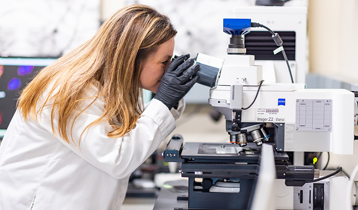
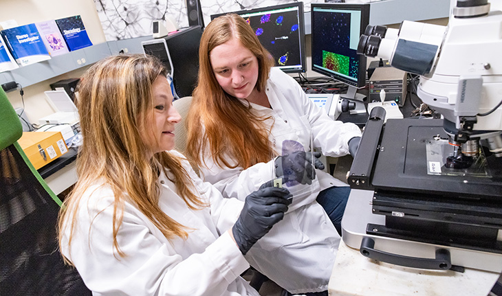
The team analyzed brain tissues as well as isolated neurons using laser capture microdissection techniques. They studied mRNA expression on a molecular level in the STG tissue and the isolated neurons. The mRNA translates the DNA code into instructions the cell machinery can recognize and use to make proteins for different body functions.
Main findings
The study identified 194 significantly different genes in the brains of people with autism. Of those genes, 143 produced more mRNA (upregulated) and 51 produced less (downregulated) in autistic brains than in typical ones.
The downregulated genes were mainly linked to brain connectivity. This may indicate that the neurons may not communicate as efficiently. Too much activity in the neurons may cause the brain to age faster in autistic individuals.
The study also found more mRNA for heat-shock proteins in autistic brains. These proteins respond to stress and activate immune response and inflammation.
“The findings from our study are really important in understanding what is happening in the brains of people with autism. Identifying these changes over time gives us an opportunity to think about some interventions that might be more useful in certain periods.”
— Cynthia Schumann, professor of neuroscience affiliated with UC Davis MIND Institute
Age-related brain differences between neurotypical and autistic people
The study identified 14 genes in bulk STG tissue that showed age-dependent differences between autistic and neurotypical individuals and three genes in isolated neurons. These genes were connected to synaptic as well as immunity and inflammation pathways.
For example, in typical brains, the expression of the HTRA2 gene is much higher before age 30 and decreases with age. In the STG neurons of people with autism, the expression levels of this gene begin lower and increase with age.
“Changes in HTRA2 have been implicated in neuronal cell loss and cell functions – such as proper protein folding, and reusing and recycling cell components,” explained co-senior author Boryana Stamova, associate professor in the Department of Neurology. She is also affiliated with the MIND Institute. “HTRA2’s role is vital for normal brain function.”
The researchers also uncovered different inflammation patterns in autistic brain tissues. Several immune and inflammation-related genes were strongly upregulated, indicating immune dysfunction that may get worse with age.
The study pointed to an age-related decrease in the gene expression involved in Gamma-aminobutyric acid (GABA) synthesis. GABA is a chemical messenger that helps slow down the brain. It works as an inhibitory neurotransmitter.
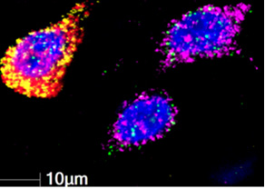
“GABA is known for producing a dampening effect in controlling neuronal hyperactivity in anxiety and stress. Our study showed age-dependent alterations in genes involved in GABA signaling in brains of people with autism,” Stamova said.
The study found direct molecular-level evidence that insulin signaling was altered in the neurons of people with autism. It also noted significant similarities of mRNA expressions in the STG region between people with autism and those with Alzheimer’s disease. These expressions may be linked to increased likelihood of neurodegenerative and cognitive decline.
“The findings from our study are really important in understanding what is happening in the brains of people with autism. Identifying these changes over time gives us an opportunity to think about some interventions that might be more useful in certain periods,” Schumann said.
Credits
The study’s co-authors are Bradley Ander of the UC Davis MIND Institute and the Department of Neurology; Alicja Omanska of the UC Davis MIND Institute and the Department of Psychiatry and Behavioral Sciences; and Michael Gandal and Pan Zhang of UCLA.
The researchers are grateful to the families of the brain donors for their invaluable gift to autism research. Brain tissues were provided by the University of Maryland Brain and Tissue Bank, Autism Tissue Program (now Autism BrainNet), Brain Endowment for Autism Research Sciences (BEARS) at the UC Davis MIND Institute, and the Harvard Brain Tissue Resource Center.
This work is supported by National Institutes of Health (NIH) grants MH108909 and the Intellectual and Developmental Disabilities Research Center at UC Davis (1U54HD079125). The study also benefited from NIH instrumentation funding (S10RR-023555, S10OD-018174) for the laser capture microdissection and RNA sequencing.
